Liver biopsy
Definition
A liver biopsy is a medical procedure performed to obtain a small piece of liver tissue for diagnostic testing. The sample is examined under a microscope by a pathologist, a doctor who specializes in the effects of disease on body tissues; in this case, to detect abnormalities of the liver. Liver biopsies are sometimes called percutaneous liver biopsies, because the tissue sample is obtained by going through the patient's skin. This is a useful diagnostic procedure with very low risk and little discomfort to the patient.
Purpose
A liver biopsy is usually done to evaluate the extent of damage that has occurred to the liver because of chronic and acute disease processes or toxic injury. Biopsies are often performed to identify abnormalities in liver tissues after other techniques have failed to yield clear results. In patients with chronic hepatitis C, liver biopsy may be used to assess the patient's prognosis and the likelihood of responding to antiviral treatment.
A liver biopsy may be ordered to diagnose or stage any of the following conditions or disorders:
- jaundice
- cirrhosis
- repeated abnormal results from liver function tests
- alcoholic liver disease
- unexplained swelling or enlargement of the liver (hepatomegaly)
- suspected drug-related liver damage such as acetaminophen poisoning
- hemochromatosis, a condition of excess iron in the liver
- intrahepatic cholestasis, the build up of bile in the liver
- hepatitis
- primary cancers of the liver such as hepatomas, cholangiocarcinomas, and angiosarcomas
- metastatic cancers of the liver (more than 20 times as common in the United States as primary cancers)
- post-liver transplant to measure graft rejection
- fever of unknown origin
- suspected tuberculosis, sarcoidosis, or amyloidosis
- genetic disorders such as Wilson's disease (a disorder in which copper accumulates in the liver, brain, kidneys, and corneas)
Demographics
According to the American Liver Foundation, liver disease affects approximately 25 million (one in 10) Americans annually. Cirrhosis accounts for over 27,000 deaths each year. Liver disease is the third most common cause of death among individuals between the ages of 25 and 59, and the seventh most common cause of all disease-related deaths.
Description
Percutaneous liver biopsy is sometimes called aspiration biopsy or fine-needle aspiration (FNA) because it is done with a hollow needle attached to a suction syringe. The special needles used to perform a liver biopsy are called Menghini or Jamshedi needles. The amount of specimen collected should be about 0.03–0.7 fl oz (1–2 cc). In many cases, the biopsy is done by a radiologist, doctor who specializes in x rays and imaging studies. The radiologist will use computed tomography (CT) scan or ultrasound to guide the needle to the target site for the biopsy. Some ultrasound-guided biopsies are performed using a biopsy gun that has a spring mechanism that contains a cutting sheath. This type of procedure gives a greater yield of tissue.
An hour or so before the biopsy, the patient will be given a sedative to aid in relaxation. The patient is then asked to lie on the back with the right elbow to the side and the right hand under the head. The patient is instructed to lie as still as possible during the procedure. He or she is warned to expect a sensation resembling a pinch in the right shoulder when the needle passes a certain nerve
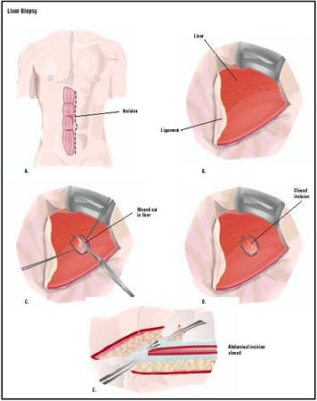
The doctor will then mark a spot on the skin of the abdomen where the needle will be inserted. The right side of the upper abdomen is thoroughly cleansed with an antiseptic solution, generally iodine. The patient is then given a local anesthetic at the biopsy site.
The doctor prepares the needle by drawing sterile saline solution into a syringe. The syringe is then attached to the biopsy needle, which is inserted into the patient's chest wall. The doctor then draws the plunger of the syringe back to create a vacuum. At this point, the patient is asked to take a deep breath and hold it. The needle is inserted into the liver and withdrawn quickly, usually within two seconds or less. The negative pressure in the syringe draws or pulls a sample of liver tissue into the biopsy needle. As soon as the needle is withdrawn, the patient can breathe normally. This step takes only a few seconds. Pressure is applied at the biopsy site to stop any bleeding and a bandage is placed over it. The liver tissue sample is placed in a cup with a 10% formalin solution and sent to the laboratory immediately. The entire procedure takes 10–15 minutes. Test results are usually available within a day.
Most patients experience minor discomfort during the procedure (up to 50% of patients), but not severe pain. According to a medical study of adult patients undergoing percutaneous liver biopsy, pain was most often described as mild to moderate (i.e., a rating of three on a scale of one to 10). Mild medications of a non-aspirin type can be given after the biopsy if the pain persists for several hours.
Diagnosis/Preparation
Liver biopsies require some preparation by the patient. Since aspirin and ibuprofen (Advil, Motrin) are known to cause excessive bleeding by inhibiting platelets and lessening clotting function, the patient should avoid taking any of these medications for at least a week before the biopsy. The doctor should check the patient's records to see whether he or she is taking any other medications that may affect blood clotting. Both a platelet count (or complete blood count ) and a prothrombin time (to assess how well the patient's blood clots) are performed prior to the biopsy. These tests determine whether there is an abnormally high risk of uncontrolled bleeding from the biopsy site, which may contraindicate the procedure. The patient should limit food or drink for a period of four to eight hours before the biopsy.
Patients should be told what to expect in the way of discomfort pre- and post-procedure. In addition, they should be advised about what medications they should not take before or after the biopsy. It is important for the clinician to reassure the patient concerning the safety of the procedure.
Before the procedure, the patient or family member must sign a consent form. The patient will be questioned about any history of allergy to the local anesthetic, and then will be asked to empty the bladder so that he or she will be more comfortable during the procedure. Vital signs , including pulse rate, temperature, and breathing rate will be noted so that the doctor can tell during the procedure if the patient is having any physical problems.
When performing the liver biopsy and blood collection that precedes it, the physician and other health care providers will follow universal precautions to maintain sterility for the prevention of transmission of blood-borne pathogens.
Some patients should not have percutaneous liver biopsies. They include those with any of the following conditions:
- a platelet count below 50,000
- a prothrombin test time greater than three seconds over the reference interval, indicating a possible clotting abnormality
- a liver tumor with a large number of veins
- a large amount of abdominal fluid (ascites)
- infection anywhere in the lungs, the lining of the chest or abdominal wall, the biliary tract, or the liver
- benign tumors (angiomas) of the liver, which consist mostly of enlarged or newly formed blood vessels and may bleed heavily
- biliary obstruction (bile may leak from the biopsy site and cause an infection of the abdominal cavity)
Aftercare
Liver biopsies are now performed as outpatient procedures in most hospitals. Patients are asked to lie on their right sides for one hour and then to rest quietly for three more hours. At regular intervals, a nurse checks the patient's vital signs. If there are no complications, the patient is discharged, but will be asked to stay in an area that is within an hour from the hospital in case delayed bleeding occurs.
Patients should arrange to have a friend or relative take them home after discharge. Bed rest for a day is recommended, followed by a week of avoiding heavy work or strenuous exercise . The patient can immediately resume eating a normal diet.
Some mild soreness in the area of the biopsy is expected after the anesthetic wears off. Irritation of the muscle that lies over the liver can also cause mild discomfort in the shoulder for some patients. Acetaminophen can be taken for minor soreness, but aspirin and ibuprofen products are best avoided. The patient should, however, call the doctor if there is severe pain in the abdomen, chest, or shoulder; difficulty breathing; or persistent bleeding. These signs may indicate that there has been leakage of bile into the abdominal cavity, or that air has been introduced into the cavity around the lungs.
Risks
The complications associated with a liver biopsy are usually minor; most will occur in the first two hours following the procedure, and greater than 95% in the first 24 hours. The most significant risk is prolonged internal bleeding. Other complications from percutaneous liver biopsies include the leakage of bile or the introduction of air into the chest cavity (pneumothorax). There is also a small chance that an infection may occur. The risk that an internal organ such as the lung, gallbladder, or kidney might be punctured is decreased when using the ultrasound- or CT-guided procedure.
Normal results
After the biopsy, the liver sample is sent to the pathology laboratory and examined. A normal (negative) result would find no evidence of pathology in the tissue sample. It should be noted that many diseases of the liver are focal and not diffuse; an abnormality may not be detected if the sample was taken from an unaffected site. If symptoms persist, the patient may need to undergo another biopsy.
The pathologist will perform a visual inspection of the sample to note any abnormalities in appearance. In cirrhosis, the sample will be fragmented and hard. Fatty liver, seen in heavy drinkers, will float in the formalin solution and will be yellow. Carcinomas are white. The pathologist will also look for deposition of bile pigments (green), indicating cholestasis (obstruction of bile flow). In preparation for microscopic examination, the tissue will be frozen and cut into thin sections, which will be mounted on glass slides and stained with various dyes to aid in identifying microscopic structures. Using the microscope, the pathologist will examine the tissue samples, and identify abnormal cells and any deposited substances such as iron or copper. In liver cancer, small dark malignant cells will be visible within the liver tissue. An infiltration of white blood cells may signal infection. The pathologist also checks for the number of bile ducts, and determines whether they are dilated. He or she also looks at the health of the small arteries and portal veins. Fibrosis will appear as scar tissue, and fatty changes are diagnosed by the presence of lipid droplets. Many different findings may be noted and a differential diagnosis (one out of many possibilities) can often be made. In difficult cases, other laboratory tests such as those assessing liver function enzymes will aid the clinician in determining the final diagnosis.
Morbidity and mortality rates
Post-biopsy complications that require hospitalization occur in approximately 1–3% of cases. Moderate pain is reported by 20% of patients, and 3% report pain severe enough to warrant intravenous pain relief. The mortality rate is approximately one in 10,000. In about 0.4% of cases, a patient with liver cancer will develop a fatal hemorrhage from a percutaneous biopsy. These fatalities result because some liver tumors are supplied with a large number of blood vessels and thus may bleed excessively.
Alternatives
Liver biopsy is an invasive and sometimes painful procedure that is also expensive (in 2002, direct costs associated with liver biopsy were $1,500–2,000). In some instances, blood tests may provide enough information to health care providers to make an accurate diagnosis and therefore avoid a biopsy. Occasionally, a biopsy may be obtained using a laparoscope (an instrument inserted through the abdominal wall that allows the doctor to visualize the liver and obtain a sample) or during surgery if the patient is undergoing an operation on the abdomen.
Resources
books
"Hepatobiliary Disorders: Introduction." In Professional Guide to Diseases, edited by Stanley Loeb, et al. Springhouse, PA: Springhouse Corporation, 2001.
Kanel, Gary C., and Jacob Korula. Liver Biopsy Evaluation, Histologic Diagnosis and Clinical Correlations. Philadelphia, PA: W.B. Saunders Company, 2000.
"Screening and Diagnostic Evaluation." In The Merck Manual of Diagnosis and Therapy, 17th Edition, edited by Robert Berkow, et al. Whitehouse Station, NJ: Merck Research Laboratories, 1999.
periodicals
Castera, Laurent, Isabelle Negre, Kamran Samii, and Catherine Buffett. "Pain Experienced during Percutaneous Liver Biopsy." Hepatology 30, no. 6 (December 1999): 1529–30.
Dienstag, Jules L. "The Role of Liver Biopsy in Chronic Hepatitis C." Hepatology 36, no. 5 (November 2002): 152–60.
Moix, F. Martin, and Jean-Pierre Raufman. "The Role of Liver Biopsy in the Evaluation of Liver Test Abnormalities." Clinical Cornerstone 3, no. 6 (2001): 13–23.
organizations
American Liver Foundation. 1425 Pompton Avenue, Cedar Grove, NJ 07009. (800) 465-4837. http://www.liverfoundation.org .
Jane E. Phillips, PhD
Stephanie Dionne Sherk
WHO PERFORMS THE PROCEDURE AND WHERE IS IT PERFORMED?
The liver biopsy requires the skill of many clinicians, including the radiologist, hepatologist, and pathologist, to make the diagnosis. Nurses will assist the physician during the biopsy procedure and in caring for the patient after the procedure. Tissues are prepared for microscopic evaluation by a histologic technician in the pathology lab. The procedure is generally performed on an outpatient basis in a hospital.
QUESTIONS TO ASK THE DOCTOR
- Why is a biopsy indicated in my case?
- How many biopsies do you perform each year? What is your rate of complications?
- What will happen when I get the results?
- What alternatives are available to me?
I had sharp pain last night that i could not sleep. So i am going to see the doctor tomorrow.otherwise during hte procedure there were no pain at all.
These medications worked in less than 5 minutes.
The Dr. used a local anesthetic at the sight of where she would be inserting the needle gun. The length of the process the doctor took using the needle gun felt like less than a minute. There was pain as penetration increased but it was bearable.
The three nurses were very kind and attentive. I was sent home with specific instructions on what not to consume. To relax. And when to notify my doctor for problems e.g. bleeding, swelling, bruising, fever.
I did experienced pain at the sight of the needle injection and took tylenol for the whole weekend.
I experienced a fever above 100 degrees about 24 hours later. I did contact the Dr. that performed the biopsy and a nurse told me to make an appointment with my primary care provider that ordered the liver biopsy.
Today (Monday) I saw a doctor with my local practice and it turns out I have strep throat.
What lead up to my liver biopsy is a liver function test showed a high alkaline phosophate level. (I take many medications and have been daily for over 20 years). My primary care physcian ordered additional blood work, which lead to an ultrasound because she was concerned about my kidney function as well. This is when a mass on my liver was found. Next was the MRI and then a liver biopsy.
I will have the results of my biopsy by the end of the week.